There is much more to your eyes than the luminous brown, green, gray, blue, or hazel orbs that look back at you in the mirror.
The inner structures of your eyes drive your ability to see clearly and are of utmost importance to your eye doctor as he or she cares for your long-term visual health.
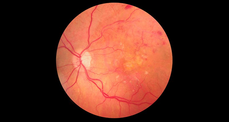
One diagnostic tool that helps your optometrist look more closely at the back of your eye and monitor eye health is digital reginal imaging (DRI).
DRI images are taken with a high-resolution digital camera to capture a detailed image of the back of your eye, providing critical information about the retina, macula and optic nerve – the eye structures that power your ability to see.
Digital retinal imaging is non-invasive and painless. You will see a bright (and we do mean bright!) flash of light as the camera takes the image, and you may see spots for a moment or two afterward. The result is a precise image of the back of your eye that your optometrist will use as a basis for long-term monitoring of your visual health. Since it’s a digital image, it is easy to store, and your optometrist can zoom in on problem areas to gain a more detailed view.
As the years go by, DRI images are a gold mine of information that can inform your future eye health. By noting any changes from earlier images, your optometrist can discover early signs of disease. That’s important, because when eye conditions are caught early, treatment is often more effective in slowing or stopping the development of symptoms.
With an up-close view of your eye’s tiny structures, your optometrist can look for abnormalities and vascular changes to the tiny capillaries that provide nutrients to your eyes and can detect a wide array of potentially sight-stealing diseases and conditions.
Here are 8 eye diseases that Digital Retinal Imaging can help diagnose and monitor:
- Glaucoma
Glaucoma, the silent thief of sight, causes damage to the optic nerve. While changes in eye pressure can indicate its early stages, patients often don’t have any noticeable symptoms until some level of vision loss has already occurred. Once nerves are damaged and vision is lost, it can’t be regained. That’s why DRI is such a powerful diagnostic tool: It can catch sight-stealing disease early when it is more treatable and can help your optometrist monitor its progression over time.
- Age-Related Macular Degeneration (AMD)
With Age-Related Macular Degeneration, the macula – that small area of the retina that provides your sharp, central vision for reading, viewing screens, and performing close-up tasks – degenerates and causes blind spots in your central vision. It can impact quality of life severely, so catching and treating it early is the key. Retinal imaging can help your optometrist see the earliest signs of AMD, such as yellowish areas called Drusen, swelling of the macula, or abnormal blood vessel growth.
- Diabetic Retinopathy
Diabetes can impact the tiny blood vessels of the eyes and cause severe vision problems. DRI empowers your optometrist to see changes to the delicate structures of the eye, including bleeding, swelling, or microaneurysms that could indicate diabetic retinopathy. With proper treatment and well-controlled blood sugar, you can slow or stop diabetic vision loss.
- Retinal Vascular Conditions
Your cells require proper nutrients to function well; when blood flow is cut off or compromised, cells and their functions suffer. That’s why monitoring the blood vessels that fuel the eye is so important. DRI can show your optometrist if there are blocked veins or arteries or if any vessels are leaking or swelling.
- Retinal Detachments and Tears
A retinal detachment is a medical emergency that must be treated quickly. Holes or tears in the retina, or its complete detachment from the back of the eye, mean that the retina is not getting the blood flow it needs. DRI can detect such tears or holes, so you can be treated promptly to prevent vision loss.
Please note: If you see dramatic changes to your visual field, such as black spots in your field of vision, or it appears that a curtain has been drawn across one eye, call us immediately or go to the emergency room if it is after hours.
- Macular Edema
Edema means swelling, and the macula is where your sharp, central vision occurs. Swelling in the macula can indicate eye diseases, including diabetes, macular degeneration, or vascular changes. Noting these changes as early as possible can help protect your vision for the future.
- Inherited Retinal Diseases
Some eye diseases, such as retinitis pigmentosa, genetic retinal dystrophies, or cone-rod dystrophy, run in families. If you have a family history of retinal disease, your optometrist will be vigilant with retinal imaging to track tissue thinning, pigment changes, or other degeneration that could indicate the early stages of this disease.
- Optic Nerve Diseases
Your optic nerve serves as the pathway for the eye’s electrical signals to get to the brain, where they are processed into what we “see.” When the optic nerve swells or atrophies, vision suffers. With a digital image of the optic nerve, your eye doctor can note changes or inconsistencies and diagnose conditions such as multiple sclerosis or inflammatory disease.
Benefits of Digital Retinal Imaging
With just a quick digital image, your optometrist can:
- Detect eye diseases and conditions in early stages.
- Plan treatments that can lead to better outcomes.
- Track disease progression and adjust treatment accordingly.
- Share images for remote consultation or second opinion when necessary.
In a flash, your eye doctor is able to accomplish so much thanks to digital retinal imaging. A painless, non-invasive photo of your eye can be the key to early detection that helps us help you enjoy the gift of sight for years to come.
If you haven’t scheduled your annual eye exam, now is a great time to call us and do so – we look forward to caring for your vision!